Fitness - FITNESS GUIDANCE : Body anatomy: Muscles guidance
Body anatomy: Muscles guidance(20 photos)
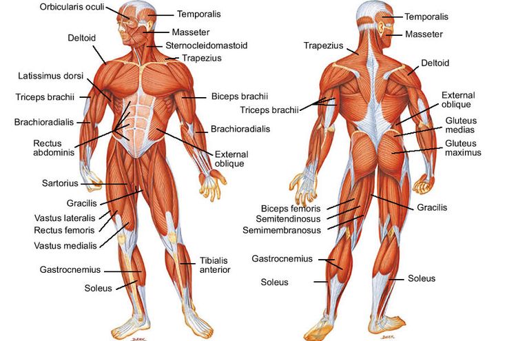
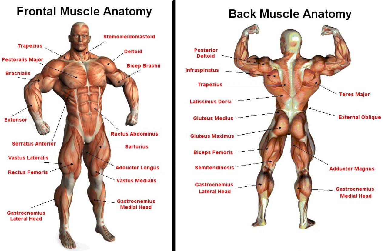
Human Muscles
Use the filters on the left to identify muscles around a particular joint as well as for specific joint actions. Click on the links below for more detailed explanations including origins, insertions and related sports injuries.
Adductor Brevis is the smallest and shortest of the three short adductor muscles. It originates on the pelvis and inserts into the thigh bone and adducts inwards and flexes the hip out forwards. It is most commonly injured in a groin strain.
Adductor Longus is the middle of the three short adductor muscles. It adducts the hip inwards and assists in hip flexion or moving the leg forwards. Originating on the ramus of the pelvis and inserts into the femur or thigh bone.
Adductor Magnus is the largest groin muscle and is one of the two long adductor muscles (gracilis is the other). It is usually described as having two parts, hamstring and adductor parts. It adducts, flexes and internally rotates the hip.
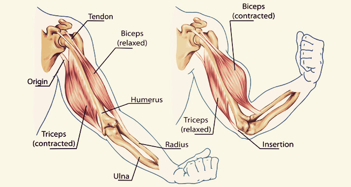
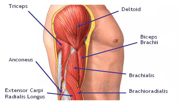
The Anconeus works alongside Triceps Brachii in extending the elbow. It also acts to pull the synovial membrane out of the way of the olecranon process when the elbow is extending.
The Biceps brachii crosses both the elbow and shoulder joints. Its action on the shoulder joint is very weak flexion. It supinates the forearm and is a strong flexor of the elbow. The bicep curl exercises is a common one to strengthen this muscle.
Biceps Femoris is one of the three muscles which form the hamstring group forming the back of the thigh. The muscle is described as having a long head (the attachment from the ischium) and a short head (attached to the femur).
The Brachialis acts to flex the elbow whether in pronation or supination, along with Biceps Brachii. As Brachialis is attached to the Ulna, which cannot rotate, it is the only true flexor of the elbow.
The erector spinae (sometimes known as sacrospinalis) is often described as a group of different muscles called iliocostalis, longissimus and spinalis.
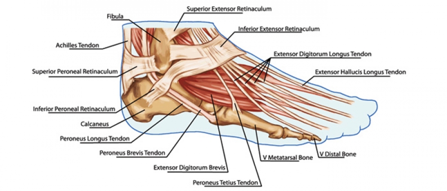
The extensor hallucis longus is the only muscle responaible for extending (pulling back) the big toe, dorsi flexes the ankle and weakly inverts the foot.
The obliques wrap around the trunk on each side to form our waists and join to the linea alba, a band of connective tissue running down the front of the abdomen.
Gluteus Medius is an important muscle in controlling the level of the hips. Weaknesses in gluteus medius often result in a trendelenburg sign, an abnormal gait cycle.
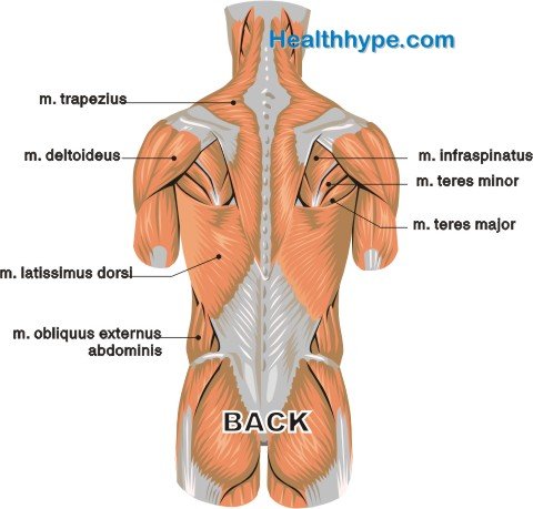
Infraspinatus is one of the rotator cuff muscles. It sits on the back of the shoulder blade, below the spine of the scapula and attaches to the greater tuberosity on the humerus.
The internal obliques wrap around the waist and insert into the linea alba, a cord like strip of connective tissue running down the centre of the abdomen.
The Latissimus dorsi muscle is one of the largest in the body. It is a powerful extensor muscle of the arm and is used extensively in chinning and climbing.
Multifidus is a series of small muscles which travel up the length of the spine. It is an important muscle in the rehabilitation of Gilmore's Groin and lower back pain.
The Pectoralis Minor muscle is the smallest of the two pectoral (chest) muscles. It works together with Serratus anterior which protracts and rotates the scapula upwards.
Popliteus is a small muscle which is often described as the key of the knee joint. It unlocks the knee joint by rotating the femur at the beginning of knee flexion to allow full knee flexion to occur.
The Rectus Femoris muscle is part of the Quadriceps muscle group. It is the only muscle of the group which crosses the hip joint and so it flexes the hip whilst extending the knee.
The Sartorius is a two joint muscle and so is weak when the knee is flexed and the hip is flexed at the same time. It works better during single movements.
Splenius is often divided into two muscles, splenius capitus (those fibres which insert on the skull) and splenius cervicis (those that insert onto the cervical transverse processes of the spine).
Subscapularis is one of the four rotator cuff muscles which cross the shoulder joint. The muscle also acts to hold the head of the humerus in position and prevents it moving forwards.
The Supraspinatus muscle is one of the four muscles which make up the rotator cuff. Its main function is to stabilise the upper arm by holding the head of the humerus in position.
Teres Minor is one of the four rotator cuff muscles surrounding the shoulder. Its main action, along with Infraspinatus is to externally rotate the shoulder joint.
Transversus Abdominis is often abbreviated to TVA. This is a very important core muscle which is vital in maintaining good posture. Activities such as Pilates focus on contraction of the TVA.
The Triceps Brachii also assists Latissimus Dorsi in extending the shoulder joint. It contracts strongly during the up phase of a push up, to straighten the arm.
Vastus Medialis is the most medially (inner) located of the quadricep muscles. The portion of the muscle just above the knee is known as VMO (vastus medialis oblique).


Human Muscles
Use the filters on the left to identify muscles around a particular joint as well as for specific joint actions. Click on the links below for more detailed explanations including origins, insertions and related sports injuries.
Adductor Brevis
Adductor Brevis is the smallest and shortest of the three short adductor muscles. It originates on the pelvis and inserts into the thigh bone and adducts inwards and flexes the hip out forwards. It is most commonly injured in a groin strain.
Adductor Longus
Adductor Longus is the middle of the three short adductor muscles. It adducts the hip inwards and assists in hip flexion or moving the leg forwards. Originating on the ramus of the pelvis and inserts into the femur or thigh bone.
Adductor Magnus
Adductor Magnus is the largest groin muscle and is one of the two long adductor muscles (gracilis is the other). It is usually described as having two parts, hamstring and adductor parts. It adducts, flexes and internally rotates the hip.
Anconeus
The Anconeus works alongside Triceps Brachii in extending the elbow. It also acts to pull the synovial membrane out of the way of the olecranon process when the elbow is extending.
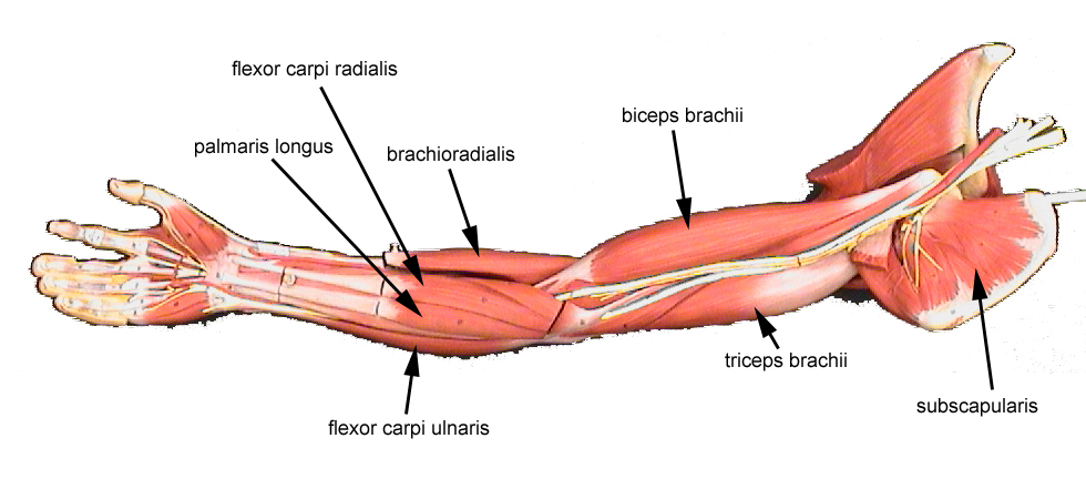
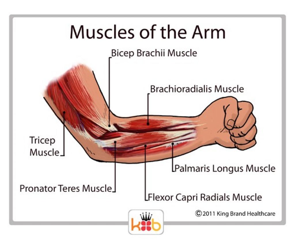
Biceps Brachii
The Biceps brachii crosses both the elbow and shoulder joints. Its action on the shoulder joint is very weak flexion. It supinates the forearm and is a strong flexor of the elbow. The bicep curl exercises is a common one to strengthen this muscle.
Biceps Femoris
Biceps Femoris is one of the three muscles which form the hamstring group forming the back of the thigh. The muscle is described as having a long head (the attachment from the ischium) and a short head (attached to the femur).
Brachialis
The Brachialis acts to flex the elbow whether in pronation or supination, along with Biceps Brachii. As Brachialis is attached to the Ulna, which cannot rotate, it is the only true flexor of the elbow.
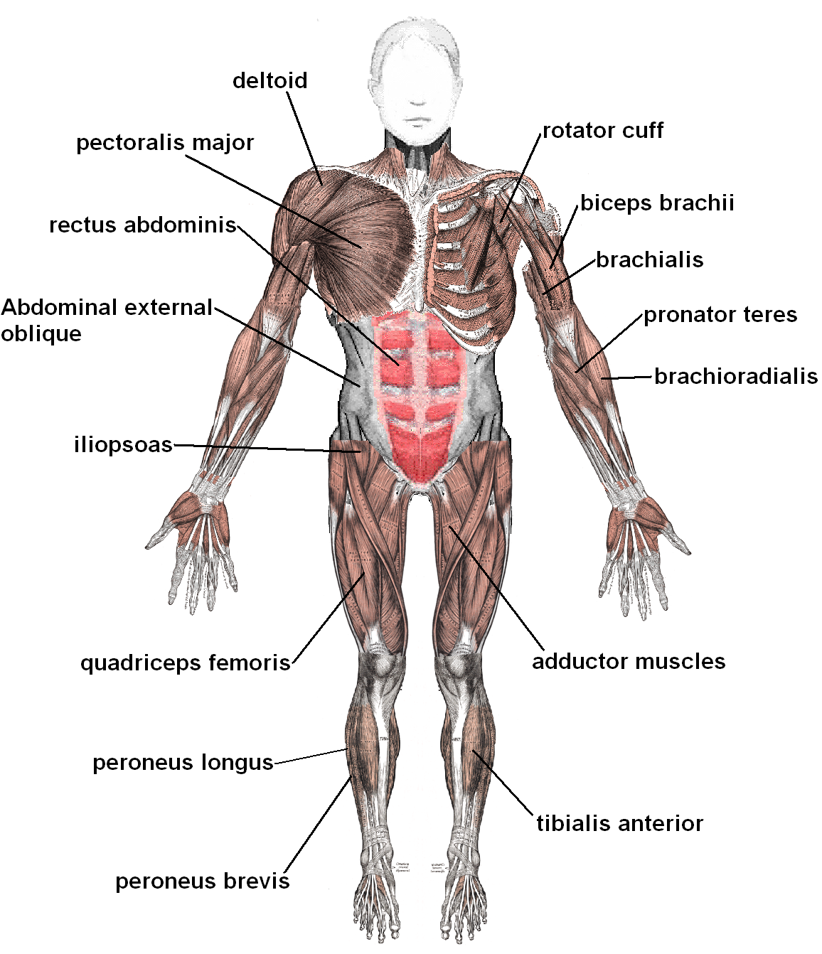
Brachioradialis
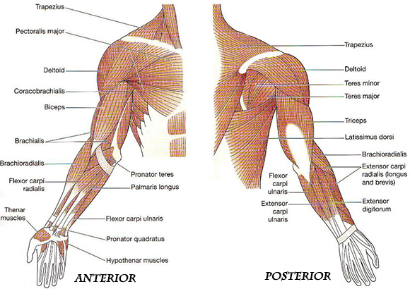
The Brachioradialis muscle flexes the elbow and supinates the forearm from a pronated position and pronates from a supinated position.
Deltoid
The deltoid muscle is used in all side lifting movements and any movement of the humerus on the scapula.
Erector Spinae
The erector spinae (sometimes known as sacrospinalis) is often described as a group of different muscles called iliocostalis, longissimus and spinalis.
Extensor Carpi Radialis Brevis
The extensor carpi radialis brevis muscle extends and abducts the wrist and is a weak extensor of the elbow.
Extensor Carpi Radialis Longus
The extensor carpi radialis longus muscle extends and abducts the wrist and is a weak extensor of the elbow.
Extensor Carpi Ulnaris
Extensor carpi ulnaris is located on the back (dorsum) of the forearm. It extends and adducts the wrist and weakly extends the elbow.
Extensor Digitorum Communis
Extensor Digitorum Longus
Extensor digitorum longus (often shortened to EDL) is found in the front of the lower leg, in the outer more muscle bound compartment.
Extensor Hallucis Longus
The extensor hallucis longus is the only muscle responaible for extending (pulling back) the big toe, dorsi flexes the ankle and weakly inverts the foot.
Extensor Policis Longus
External Obliques
The obliques wrap around the trunk on each side to form our waists and join to the linea alba, a band of connective tissue running down the front of the abdomen.
Flexor Carpi Radialis
The flexor carpi radialis muscle flexes and abducts the wrist and is a weak flexor of the elbow.
Flexor Carpi Ulnaris
The flexor carpi ulnaris muscle flexes and adducts the wrist as well as being a weak flexer of the elbow.
Flexor Digitorum Longus
Flexor Digitorum Longus causes the toes to grip and mold to the floors surface which is vital in maintaining balance on rough surfaces.
Flexor Digitorum Superficialis
The flexor digitorum superficialis muscle flexes or bends the fingers, flexes the wrist and is a weak flexor of the elbow.
Flexor Hallucis Longus
Flexor Hallucis Longus bends the big toe when you curl up your foot. It is called 'Hallucis' as the word Hallux means great or big toe in latin.
Flexor Pollicis Longus
The flexor pollicis longus muscle lfexes the thumb and wrist and is a weak flexor of the elbow.
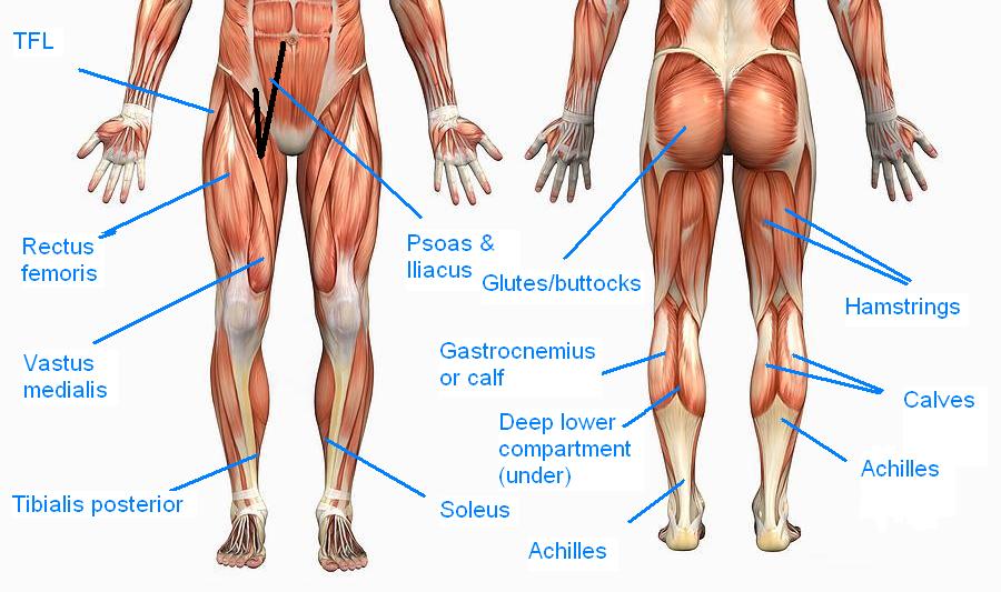
Gastrocnemius
Gastrocnemius muscle is a strong plantar flexor of the ankle and weakly flexes the knee.
Gluteus Maximus
Gluteus Maximus is the largest and most superficial of the three gluteal muscles which forms the rounded shape of the buttocks.
Gluteus Medius
Gluteus Medius is an important muscle in controlling the level of the hips. Weaknesses in gluteus medius often result in a trendelenburg sign, an abnormal gait cycle.
Gluteus Minimus
This is the smallest of the three gluteal muscles. It abducts the hip and assists with internal rotation as the femur abducts.
Gracilis
Gracilis is another muscle which works in conjunction with the groin muscles and is also a weak knee flexor.
Groin Muscles
The groin muscles are sometimes also call the 'adductor's. This describes the movement that they all perform. There are five adductors in total.
Hamstrings
The hamstring muscles are found at the back of the thigh. They are three muscles which act on both the hip and knee joints.
Iliopsoas
A powerful hip flexor, also assists in externally rotating the femur.
Infraspinatus
Infraspinatus is one of the rotator cuff muscles. It sits on the back of the shoulder blade, below the spine of the scapula and attaches to the greater tuberosity on the humerus.
Internal Obliques
The internal obliques wrap around the waist and insert into the linea alba, a cord like strip of connective tissue running down the centre of the abdomen.
Latissimus Dorsi
The Latissimus dorsi muscle is one of the largest in the body. It is a powerful extensor muscle of the arm and is used extensively in chinning and climbing.
Levator Scapulae
Shrugging the shoulders (scapula elevation) requires the use of levator scapulae and Trapezius.
Multifidus
Multifidus is a series of small muscles which travel up the length of the spine. It is an important muscle in the rehabilitation of Gilmore's Groin and lower back pain.
Pectineus
Pectineus is positioned between the Iliopsoas and Adductor Longus muscles and is part of the short adductor group with adductors brevis and longus.
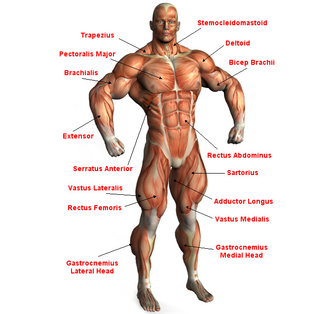
Pectoralis Major
Pectoralis major is the largest and most superficial of the chest muscles, Its action depends on the position of the arm.
Pectoralis minor
The Pectoralis Minor muscle is the smallest of the two pectoral (chest) muscles. It works together with Serratus anterior which protracts and rotates the scapula upwards.
Peroneus Brevis
Peroneus Brevis everts (turn outwards) the foot and plantar flex the ankle.
Peroneus Longus
Peroneus Longus is one of the peroneals muscle group which pass down the outside of the lower leg and evert (turn out) the foot.
Piriformis
The Piriformis muscle is an important muscle. The sciatic nerve passes underneath this muscle on its route down to the posterior thigh.
Popliteus
Popliteus is a small muscle which is often described as the key of the knee joint. It unlocks the knee joint by rotating the femur at the beginning of knee flexion to allow full knee flexion to occur.
Pronator Quadratus
Pronator Quadratus works in conjunction with Triceps Brachii during pronation with elbow extension.
Pronator Teres
Pronator Teres works the hardest when the elbow is flexing and the hand simultaneously pronating.
Quadratus Lumborum
The quadratus lumborum or QL is a common cause of back pain which is to one side and comes on after lifting or twisting.
Quads (Quadriceps Muscles)
Quads (Quadriceps Muscles)
Rectus Abdominis
Rectus Abdominis is the most superficial of the abdominal muscles. It is this muscle which forms the six-pack shape!
Rectus Femoris
The Rectus Femoris muscle is part of the Quadriceps muscle group. It is the only muscle of the group which crosses the hip joint and so it flexes the hip whilst extending the knee.
Rhomboids
There are two rhomboid muscles - Rhomboid Major and Rhomboid Minor. Rhomboid major is larger and positioned below rhomboid minor.
Sartorius
The Sartorius is a two joint muscle and so is weak when the knee is flexed and the hip is flexed at the same time. It works better during single movements.
Semimembranosus
Semimembranosus is the most medial of the three hamstring muscles. It extends and internally rotates the hip and flexes the knee.
Semitendinosus
The semitendinosus muscle extends and internally rotates the hip as well as flexing the knee.
Serratus Anterior
The Serratus Anterior muscle is used in activities which draw the scapula forwards. It is used strongly in push-ups and bench presses.
Shoulder Girdle
Soleus
Soleus is a large large muscle, deep to Gastrocnemius. Together the Gastrocnemius, Soleus and Plantaris are known as Triceps Surae.
Splenius
Splenius is often divided into two muscles, splenius capitus (those fibres which insert on the skull) and splenius cervicis (those that insert onto the cervical transverse processes of the spine).
Sternocleidomastoid
Sternocleidomastoid (SCM) can clearly be seen when you turn your head to one side, on the opposite side of the neck.
Subscapularis
Subscapularis is one of the four rotator cuff muscles which cross the shoulder joint. The muscle also acts to hold the head of the humerus in position and prevents it moving forwards.
Supinator
The Supinator muscle assists Biceps Brachii in supinating the hand, that is turning it over so that the palm faces up.
Supraspinatus
The Supraspinatus muscle is one of the four muscles which make up the rotator cuff. Its main function is to stabilise the upper arm by holding the head of the humerus in position.
Tensor Fascia Latae
The Tensor Fasciae Latae (TFL) is a small muscle which attaches inferiorly to the long thick strip of fascia, known as the iliotibial band (ITB).
Teres Major
Teres Minor is one of the four rotator cuff muscles surrounding the shoulder. Its main action, along with Infraspinatus is to externally rotate the shoulder joint.
Teres Minor
The Glutes
The 'Glutes' is an abbreviation of the gluteals - also known as the buttock muscles. The three main ones are the Gluteus Maximus, Medius and Minimus.
Tibialis Anterior
Tibialis anterior forms the main fleshy part of the outside of the shin. It is a dorsiflexor of the ankle.
Tibialis Posterior
The Tibialis Posterior is the deepest of all the calf muscles. It helps to support the arch of the foot.
Transversus Abdominis
Transversus Abdominis is often abbreviated to TVA. This is a very important core muscle which is vital in maintaining good posture. Activities such as Pilates focus on contraction of the TVA.
Trapezius Muscle
The trapezius muscle (Trapz) is a large muscle consisting of four parts covering the upper back, shoulders and neck.
Triceps Brachii
The Triceps Brachii also assists Latissimus Dorsi in extending the shoulder joint. It contracts strongly during the up phase of a push up, to straighten the arm.
Vastus Intermedius
Vastus Intermedius is one of four quadricep muscles, located deep in the thigh underneath the Rectus Femoris muscle.
Vastus lateralis
Vastus Lateralis is the most lateral (outer) of the four quadriceps muscles and is felt on the outside top of the thigh.
Vastus Medialis
Vastus Medialis is the most medially (inner) located of the quadricep muscles. The portion of the muscle just above the knee is known as VMO (vastus medialis oblique).
» Read more
Tune Me In Now on guidance to identify each muscle in a human body *** CLICK AT ANY PICTURE TO ENLARGE IT and see a SLIDE SHOW***